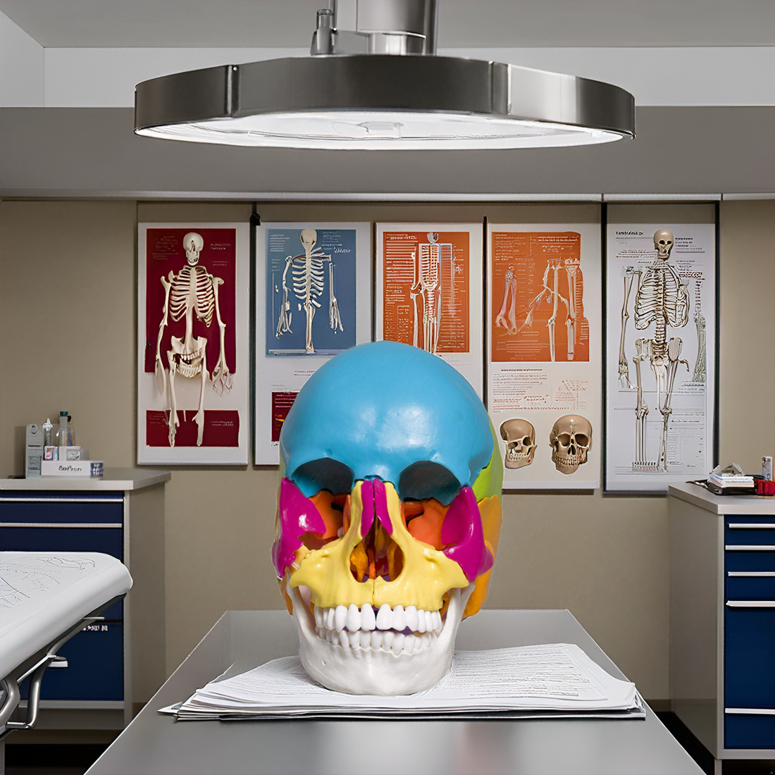
Overview of Skull Anatomy
The skull is a complex, protective structure made of bone. It is essential for safeguarding the brain and supporting various sensory functions. The skull anatomy is divided into two main sections: the cranium, which encloses and shields the brain, and the facial bones, including the mandible or jawbone, that shape the face. In scientific terms, the part that encases the brain is known as the neurocranium. At the same time, the viscerocranium forms the facial skeleton, including the jaw
Beyond its role in protecting the brain, the skull supports vital sensory organs like the eyes, ears, nose, and mouth. It ensures that the eyes are positioned correctly for optimal vision and secures the placement of the ears to assist in hearing and determining the direction of sound. Additionally, the skull aids breathing and eating by housing the nasal cavity and jaw.
The skull’s intricate design ensures protection and functionality, playing a central role in survival by enabling sensory perception and environmental interaction.
Skull Anatomy Labeled Diagram
Parts of Skull
Cranial Bones (8 bones)
- Frontal Bone
- Parietal Bones (2)
- Temporal Bones (2)
- Occipital Bone
- Sphenoid Bone
- Ethmoid Bone
Facial Bones (14 bones)
- Nasal Bones (2)
- Maxillae (2) & Mandible
- Zygomatic Bones (2)
- Lacrimal Bones (2)
- Palatine Bones (2)
- Inferior Nasal Conchae (2)
- Vomer
Additional Structures
- Sutures
· Coronal Suture
· Sagittal Suture
· Lambdoid Suture
· Squamous Suture
- Foramina
· Foramen Magnum
· Optic Foramen
Human Skull Anatomy: Parts & Functions
Cranial Bones (8 bones)
The cranium, or neurocranium, consists of eight bones that cover and safeguard the brain. It is divided into two main parts: the cranial roof and the cranial base.
The cranial roof, known as the calvarium, includes the frontal, occipital, and parietal bones. The cranial base comprises all eight cranial bones. These protect the brain and support structures of the face.

1. Frontal Bone
The frontal bone has two main parts: the squamous part and the orbital part. The squamous part is the large, flat area that makes up most of the forehead.
The orbital part is smaller and lies horizontally, forming the top of the eye sockets and helping shape the nasal cavities.
Sometimes, there is a third part called the nasal part, which connects the brow ridges to the nasal bones below and the bones on the sides of the nose (lacrimal and maxilla).
The key function of the frontal bone is to shape the forehead and protect the brain’s frontal lobe. It also helps form the eye sockets and the front of the brain cavity.
2. Parietal Bones (2)
The parietal bones are paired structures on each side of the skull. It forms the upper and side portions of the head. They rest above the brain’s parietal lobes and are shielded by a fibrous tissue called the epicranial aponeurosis.
These bones are part of the neurocranium—the part of the skull that houses and protects the brain—alongside other key bones like the frontal, ethmoid, sphenoid, temporal, and occipital bones. The parietal bones mainly shape the top of the skull, with a smaller section contributing to the skull base.
Their primary function is to safeguard the brain from injury. Slightly curved and rectangular, each parietal bone has two surfaces, four borders, and four corners.
These borders link with other skull bones at junctions called cranial sutures. The inner surfaces of the parietal bones feature grooves and depressions that accommodate blood vessels and other essential structures.![]()
3. Temporal Bones (2)
The temporal bones are a pair of bones on either side of the skull. It forms parts of both the base and the sides of the head. They have a complex shape because they anchor various muscles and connect with neighboring bones.
These bones are also crucial passageways for nerves and blood vessels moving in and out of the skull. Hidden within the temporal bones are the essential structures that control hearing and balance, including portions of the middle and inner ear.
From the back of the skull, the temporal bones are visible on the sides, with the mastoid process—a noticeable, rounded bump—standing out.
The temporal bone comprises several distinct sections:
1. The broad,
2. Flat squamous part
3. The pyramid-shaped petrous part
4. The tympanic part, which surrounds the ear canal
5. The slender styloid process is a pointed projection beneath the ear.
4. Occipital Bone
The occipital bone is located at the back and base of the skull. It is a crucial structure that protects the brain’s occipital lobes.
The bone is curved trapezoid and has a key feature called the foramen magnum. This large oval opening allows the spinal cord to connect with the brain.
The occipital bone is divided into three distinct sections based on its features.
1. The front section, the basilar part or basioccipital, is a thick, rectangular segment extending forward.
2. On either side of the foramen magnum lie the lateral parts, known as the exoccipitals, which support the skull’s connection to the spine.
3. The back section, or squamous part, forms the largest and most curved area, supporting the skull’s rear.
This design plays a vital role in brain protection and the connection between the brain and spinal cord. It makes it essential for structural integrity and neurological function.
5. Sphenoid Bone
The sphenoid bone is one of the most intricate bones in the human body. Because of its unique shape, it is sometimes called the “wasp bone.”
It forms a large part of the base of the skull, especially in the middle, and helps create the floor of the middle section of the skull’s interior.
This bone is closely connected to important soft tissues like the cranial nerves and parts of the brain. Its main role is to provide openings and pathways (foramina and canals) for nerves and blood vessels to pass in and out of the skull.

6. Ethmoid Bone
Facial Bones (14 bones)
The cranium has two main parts: the neurocranium and the viscerocranium. The neurocranium protects the brain, while the viscerocranium, also called the facial skeleton, shapes our face.
The facial skeleton has 14 bones.
- Six of them come in pairs, with one on each side of the face. These paired bones are the inferior nasal conchae, nasal bones, maxillae (upper jaw), palatine bones, lacrimal bones, and zygomatic bones (cheekbones).
- There are also two single bones: the mandible (lower jaw) and the vomer.
Besides giving shape to the face, the viscerocranium protects important organs like the eyes and mouth. It has spots for muscles to attach and tiny holes, called foramina, that let nerves and blood vessels pass through.
These shape the face and provide cavities for the sense organs (eyes, nose, and mouth).
